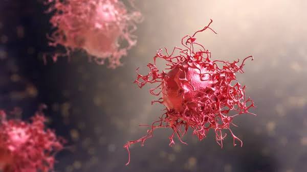
Neoplasia?
Topics cover.
- What is Neoplasia?
- What is benign & malignant tumor?
- Classification & nomenclature of neoplastic disease?
- Chemical, physical, & biological carcinogenesis?
- Clinical & gross structure of Dysplasia?
- What is Invasion & Metastatis?
- Cytological features of malignancy?

What is Neoplasia?
Neoplasia is the abnormal and uncontrolled growth of cells in the body, leading to the formation of a tumor or mass. These cells, called neoplastic cells, often lack the typical regulatory mechanisms that control cell growth. Neoplasia can be benign (non-cancerous) or malignant (cancerous), with the latter having the potential to invade nearby tissues and spread to other parts of the body.

What is benign & malignant tumor?
Benign Tumor
A benign tumor is a non-cancerous growth of cells that does not invade nearby tissues or spread to other parts of the body. While it can grow and cause health issues if it presses against surrounding structures, it usually remains localized. Benign tumors are typically not life-threatening and can often be removed through surgery if necessary.
Malignant Tumor (Cancer)
A malignant tumor, also known as cancer, is a growth of abnormal cells that can invade nearby tissues and spread to distant parts of the body through a process called metastasis. Malignant cells have the ability to invade blood vessels and lymphatic channels, allowing them to migrate to other organs and form secondary tumors (metastases). Malignant tumors can be life-threatening if not treated and managed effectively. They require various treatment approaches, including surgery, chemotherapy, radiation therapy, and targeted therapies.
Classification and nomenclature of neoplastic disease?
Neoplastic diseases, which involve the abnormal growth of cells leading to the formation of tumors, are classified and named based on various criteria. The classification and nomenclature often consider factors such as the tissue of origin, cellular characteristics, behavior (benign or malignant), and histological appearance. Here's an overview:
1. Tissue of Origin:
- Neoplastic diseases can be classified based on the tissue or organ from which they originate. For example, "breast cancer" originates in breast tissue, and "lung cancer" originates in lung tissue.
2. Benign vs. Malignant:
- Tumors are classified as benign or malignant based on their behavior.
- Benign tumors are non-cancerous and do not invade nearby tissues or spread to distant sites.
- Malignant tumors are cancerous, with the potential to invade surrounding tissues and metastasize to other parts of the body.
3. Histological Appearance:
- Pathologists often classify tumors based on their microscopic appearance (histology). This includes factors such as cell size, shape, and tissue architecture.
- Examples include adenoma (benign glandular tumor) and adenocarcinoma (malignant glandular tumor).
4. Cell Type and Differentiation:
- Tumors may be classified by the type of cells involved and their level of differentiation (how closely they resemble normal cells).
- Well-differentiated tumors closely resemble normal tissue, while poorly differentiated or undifferentiated tumors have more abnormal-looking cells.
5. Molecular Characteristics:
- Advances in molecular biology have led to the classification of tumors based on specific genetic or molecular alterations.
- For example, some breast cancers are classified as "HER2-positive" based on the overexpression of the HER2 gene.
6. Staging and Grading:
- Malignant tumors are often staged to determine the extent of disease, which guides treatment decisions. Staging considers factors such as tumor size, lymph node involvement, and distant metastasis.
- Grading assesses the degree of cellular differentiation and helps predict tumor behavior.
7. International Classification Systems:
- International classification systems like the TNM (Tumor, Node, Metastasis) staging system and the World Health Organization (WHO) classification provide standardized frameworks for categorizing and naming neoplastic diseases.
In practice, neoplastic diseases are typically named based on a combination of these factors. For example, "well-differentiated squamous cell carcinoma of the lung" describes a malignant lung tumor with squamous cell characteristics that are well-differentiated (resembling normal cells to some extent). Accurate classification and nomenclature are essential for diagnosis, treatment planning, and research into these diseases.
Chemical, physical, biological carcinogenesis?
Carcinogenesis, the process by which normal cells transform into cancer cells, can occur through multiple mechanisms, including chemical, physical, and biological factors. Here's an overview of each type of carcinogenesis:
1. Chemical Carcinogenesis:
Definition: Chemical carcinogenesis involves the induction of cancer by exposure to specific chemicals or compounds. These chemicals are called carcinogens.
- Examples:Carcinogens can include tobacco smoke, certain industrial chemicals, pesticides, and naturally occurring substances like aflatoxins produced by molds.
- Mechanism: Chemical carcinogens can damage DNA, leading to mutations in critical genes that regulate cell growth and division. This can result in the uncontrolled growth of cells and the development of cancer.
- Prevention:Reducing exposure to known carcinogens, such as quitting smoking or using protective equipment in workplaces with chemical hazards, can help prevent chemical carcinogenesis.
2. Physical Carcinogenesis:
- Definition:Physical carcinogenesis involves the induction of cancer by exposure to physical agents, such as radiation or chronic physical irritation.
- Examples:Ionizing radiation (e.g., X-rays, gamma rays), ultraviolet (UV) radiation from the sun, and asbestos fibers are physical carcinogens.
- Mechanism: Ionizing radiation can directly damage DNA, while UV radiation can cause mutations and skin cancers. Chronic irritation can lead to cell turnover and increased risk of mutations.
- Prevention: Protecting oneself from excessive sun exposure, following safety protocols in radiation-related work, and avoiding asbestos exposure can help reduce the risk of physical carcinogenesis.
3. Biological Carcinogenesis:
- Definition: Biological carcinogenesis involves the induction of cancer by infectious agents, such as viruses or certain bacteria.
- Examples: Human papillomavirus (HPV) is linked to cervical cancer, hepatitis B and C viruses are associated with liver cancer, and Helicobacter pylori is linked to stomach cancer.
- Mechanism:These infectious agents can introduce genetic changes or cause chronic inflammation, leading to cellular damage and increased cancer risk.
- Prevention:Vaccines against certain viruses (e.g., HPV, hepatitis B) and early treatment of infections can reduce the risk of biological carcinogenesis.
It's important to note that carcinogenesis is a complex and multifactorial process often involving a combination of these mechanisms. Additionally, individual susceptibility and genetic factors play a role in determining an individual's risk of developing cancer. Cancer prevention efforts often focus on reducing exposure to known carcinogens, promoting healthy lifestyles, and early detection through screening and vaccination.
.jpeg)
Clinical and Gross structure of Dysplasia?
Dysplasia is a term used in pathology to describe abnormal changes in the structure and organization of cells within a tissue. Dysplasia can be a pre-cancerous condition, indicating an increased risk of developing cancer in the affected tissue. It can occur in various tissues throughout the body. Here's an overview of the clinical and gross structure aspects of dysplasia:
Clinical Aspect:
- Dysplasia is often detected through routine medical examinations or screenings, such as Pap smears for cervical dysplasia.
- Symptoms may not be noticeable in early stages, and dysplasia is typically identified through microscopic examination of tissue samples (biopsy).
Gross Structure:
- Grossly, dysplasia may not present any visible or palpable changes to the naked eye. In many cases, the tissue appears normal in color, size, and texture.
- Dysplasia is primarily characterized by abnormal cellular changes when examined microscopically. These changes may include:
- Altered cell size and shape: Dysplastic cells can vary in size and shape, with some appearing larger or smaller than normal.
- Increased nuclear-to-cytoplasmic ratio: Dysplastic cells often have a larger nucleus compared to the surrounding cytoplasm, indicating abnormal cell growth.
- Loss of normal tissue organization: Dysplastic cells may lose their normal arrangement and organization within the tissue.
- Variation in nuclear morphology: Dysplastic nuclei may show irregular shapes, size variation, and increased chromatin (genetic material) density.
- Hyperchromasia: Dysplastic nuclei may appear darker (hyperchromatic) due to increased DNA content.
- Abnormal mitotic figures: Dysplastic tissue may exhibit an increased number of abnormal cell divisions (mitotic figures).
It's important to note that dysplasia can vary in severity, ranging from mild (low-grade) to moderate (intermediate-grade) to severe (high-grade). Higher-grade dysplasia is associated with a greater risk of progressing to cancer.
Dysplasia is a concerning finding because it suggests a heightened risk of cancer development in the affected tissue. Medical management typically involves close monitoring, surveillance, or interventions, such as removal of dysplastic tissue to prevent progression to cancer. The specific approach depends on the location and severity of the dysplasia. Early detection and management of dysplasia are crucial for preventing the development of invasive cancer.
What is Invasion and metastatis?
Invasion and metastasis are key processes in the progression of cancer, describing how cancer cells spread from their original site to other parts of the body. These processes are crucial in understanding the malignant nature of cancer:
1. Invasion:
Definition: Invasion refers to the ability of cancer cells to penetrate and invade nearby tissues surrounding the primary tumor site.
-Mechanism:Cancer cells acquire the ability to break through the basement membrane (a barrier that separates different layers of tissues) and invade the neighboring tissues, such as adjacent organs or blood vessels.
- Local Impact:Invasion can lead to destruction of normal tissues, organ dysfunction, and local symptoms at the primary tumor site.
- Clinical Implication: Invasion is a hallmark of malignancy and contributes to the ability of cancer to cause harm locally.
2. Metastasis:
- Definition: Metastasis is the process by which cancer cells break away from the primary tumor, enter the bloodstream or lymphatic system, and spread to distant sites in the body where they form secondary tumors (metastatic tumors).
-Mechanism: Metastatic cancer cells can survive the journey through the bloodstream or lymphatic system, exit at a distant location, and establish new tumor growth.
- Distant Impact: Metastasis can affect various organs and tissues, leading to the formation of tumors in distant sites from the primary tumor.
- Clinical Implication:Metastasis is a major factor contributing to the high mortality associated with cancer. It often makes cancer challenging to treat, as it requires addressing tumors at multiple sites.
Metastatic Cascade:
The process of metastasis involves several stages, including intravasation (entry into blood or lymphatic vessels), circulation through the bloodstream or lymphatic system, extravasation (exit from vessels), and colonization at a distant site. Only a small fraction of cancer cells that enter the circulation successfully complete this complex journey to form metastatic tumors.
Understanding invasion and metastasis is crucial in cancer diagnosis, staging, and treatment planning. Therapies aimed at preventing or treating metastasis are a significant focus of cancer research and treatment strategies. Early detection and intervention are key to improving outcomes for individuals with cancer.
Cytological features of malignancy?
Cytological features, as observed in laboratory examinations of cells, can provide important clues about the presence of malignancy (cancer). Cytological evaluation involves the examination of cell samples, typically obtained through procedures like fine needle aspiration (FNA) or Pap smears. Here are some common cytological features suggestive of malignancy:
1. Abnormal Cell Size and Shape:
- Malignant cells may vary in size and shape, displaying anisocytosis (unequal cell size) and anisokaryosis (unequal nuclear size).
2. Increased Nuclear-to-Cytoplasmic Ratio (N/C Ratio):
- Malignant cells often have a higher N/C ratio compared to normal cells. This means that the nucleus is relatively larger compared to the surrounding cytoplasm.
3. Nuclear Abnormalities:
- Pleomorphism: Malignant cells may show pleomorphism, which means they have irregular or varying nuclear shapes.
- Hyperchromasia: The nucleus of malignant cells may appear darker due to increased chromatin (genetic material) density.
- Irregular Nuclear Borders: The nuclear membrane may be irregular or show irregular contours.
4. Increased Mitotic Activity:
- Malignant cells frequently undergo more frequent and abnormal cell divisions (mitotic figures) compared to normal cells.
5. Loss of Tissue Organization:
- Malignant cells may lose the normal organization and orientation seen in healthy tissues. They often appear disorganized and lack the characteristic patterns of normal tissue.
6. Abnormal Cell Clusters:
- Malignant cells may form irregular clusters or groups, in contrast to the organized arrangement of normal cells.
7. Inflammation and Necrosis:
- Areas of inflammation and necrosis (cell death) may be present in malignant samples due to the rapid and chaotic growth of cancer cells.
8. Cellular Atypia:
- Cellular atypia refers to abnormal cell structure and morphology, which is often indicative of malignancy.
9. Increased Nucleoli:
- Nucleoli (small, round structures within the nucleus) may be prominent and numerous in malignant cells.
10. Background Changes:
- The background of a cytological sample may contain abnormal cell debris, inflammatory cells, or blood.
It's important to note that while these features can suggest malignancy, a definitive diagnosis typically requires further evaluation, including histological examination (examination of tissue sections) and potentially genetic or molecular testing. Additionally, not all features may be present in every case of malignancy, and some benign conditions may exhibit cytological atypia. Accurate diagnosis and clinical management should be performed by qualified pathologists and healthcare professionals.
.jpeg)

.jpeg)
Soy Richard, estoy aquí para testificar sobre un gran herbolario que curó a mi esposa del cáncer de mama. Su nombre es Dr. Imoloa. Mi esposa pasó por este dolor durante 3 años, yo casi gasté todo lo que tenía, hasta que vi algunos testimonios en línea sobre cómo el Dr. Imoloa los curó de sus enfermedades, inmediatamente lo contacté. Luego me dijo las cosas necesarias que debía hacer antes de enviar la medicina herbaria. Ojalá lo hubiera hecho a través del servicio de mensajería DHL y nos instruyó sobre cómo aplicar o beber el medicamento durante dos semanas. y para gran sorpresa, antes de la tercera semana superior mi esposa se sintió aliviada de todos los dolores. Créanme, así fue como mi esposa fue curada del cáncer de mama por este gran hombre. También tiene poderosas medicinas a base de hierbas para curar enfermedades como: enfermedad de Alzheimer, enfermedad de Parkinson, cáncer vaginal, epilepsia, trastornos de ansiedad, enfermedades autoinmunes, dolor de espalda, esguince de espalda, trastorno bipolar, tumor cerebral, maligno, bruxismo, bulimia, enfermedad del disco cervical, cardiovascular. Enfermedades, Neoplasias, Enfermedad respiratoria crónica, Trastorno mental y del comportamiento, Fibrosis quística, Hipertensión, Diabetes, Asma, Artritis autoinmune media inflamatoria ed. enfermedad renal crónica, enfermedad inflamatoria de las articulaciones, impotencia, espectro alcohólico feta, trastorno distímico, eccema, tuberculosis, síndrome de fatiga crónica, estreñimiento, enfermedad inflamatoria intestinal, enfermedad de lupus, úlcera bucal, cáncer de boca, dolor corporal, fiebre, hepatitis ABC, sífilis, diarrea, VIH/SIDA, enfermedad de Huntington, acné de espalda, insuficiencia renal crónica, enfermedad de Addison, dolor crónico, dolor de Crohn, fibrosis quística, fibromialgia, enfermedad inflamatoria intestinal, enfermedad fúngica de las uñas, enfermedad de Lyme, enfermedad de Celia, linfoma, depresión mayor, maligno melanoma, Manía, Melorreostosis, Enfermedad de Meniere, Mucopolisacaridosis, Esclerosis múltiple, Distrofia muscular, Artritis reumatoide. Puede comunicarse con él por correo electrónico a través de drimolaherbalmademedicine@gmail.com
ReplyDelete