Posts

Showing posts from May, 2023

Platelet's related disease

Image
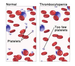
What is platelet disease? Platelet illness alludes to a gathering of issues portrayed by irregularities in the construction, capability, creation, or obliteration of platelets. These problems can bring about weakened platelet capability, decreased platelet count (thrombocytopenia), or strange platelet morphology. Why it's happens?  Platelet infections can be gained or acquired. Obtained platelet problems might happen because of different variables, like drugs, contaminations, immune system conditions, or fundamental illnesses influencing the bone marrow or insusceptible framework. Acquired platelet issues are normally hereditary circumstances that are available from birth and are brought about by changes in qualities engaged with platelet creation or capability. A few instances of platelet problems include: 1. Thrombocytopenia: A condition portrayed by a low platelet count, which can prompt expanded draining propensities. 2. Von Willebrand Sickness: An acquired draining problem br

Platelet-structure, function, formation, types?

Image
What is platelet? Platelets, also known as thrombocytes, are small, disc-shaped cells found in the blood. They are formed in the bone marrow from large cells called megakaryocytes. Platelets play a crucial role in the process of blood clotting or hemostasis. Structure of platelet Activation of platelet?  Platelets can be actuated by different upgrades, including: 1. Endothelial harm: When veins are harmed, the fundamental endothelial cells are uncovered. Platelets perceive and stick to the harmed endothelium, starting the enactment interaction. 2. Thrombin: Thrombin is a critical compound in the thickening fountain. It is created because of the coagulation interaction and fills in as a powerful platelet activator. Thrombin ties to explicit receptors on platelets, setting off their enactment. 3. ADP (Adenosine Diphosphate): ADP is set free from platelets themselves or from harmed tissues. It goes about as a flagging particle, restricting to platelet surface receptors and prompting pla

Cellular injury?

Image
  What is cellular injury?  Cellular injury refers to the damage or harm caused to cells due to various factors, such as physical trauma, chemical exposure, infection, or lack of oxygen. It disrupts the normal functioning of cells and can lead to cell death or dysfunction. Common mechanisms of cellular injury include oxidative stress, inflammation, and disruption of cellular structures. Understanding cellular injury is crucial in studying diseases and developing treatments to mitigate or prevent damage to cells. Types of cellular injury Cell injury can happen because of different factors and can appear in changed structures. Here are a few normal kinds of cell wounds: 1. Hypoxic Injury:  It results from diminished oxygen supply to the cells. It very well may be brought about by elements like ischemia (diminished blood stream), respiratory illnesses, or insufficient oxygenation of the blood. Hypoxic injury can prompt cell brokenness and, if extreme and delayed, can bring about cell demi

Activation of T lymphocytes

Image
Figure - ACTIVATION OF T LYMPHOCYTES  Why T lymphocytes activation is important The activation of T lymphocytes is essential for initiating and coordinating the adaptive immune response.  T lymphocytes, also known as T cells, play a crucial role in recognizing and eliminating infected cells, cancer cells, and other foreign substances in the body. The activation of T cells requires the following requirements: 1. Antigen Presentation: T lymphocytes recognize antigens, which are small fragments of proteins derived from pathogens or abnormal cells. Antigens are presented to T cells by antigen-presenting cells (APCs), primarily dendritic cells. APCs process antigens and display them on their surface using a specialized molecule called the major histocompatibility complex (MHC). 2. T Cell Receptor (TCR) Recognition: T lymphocytes possess a unique T cell receptor (TCR) on their surface that can specifically recognize the antigens presented by APCs. The TCR interacts with the antigen-MHC compl

Colorimeter

Image
  What is colorimeter?  A colorimeter is a device used to measure the intensity of color in a solution or sample. It quantitatively determines the concentration of a solute by measuring the absorbance or transmittance of light at specific wavelengths. The colorimeter consists of a light source, a filter or monochromator to select a specific wavelength of light, a sample chamber, and a detector to measure the intensity of light that passes through the sample. Here's how a colorimeter typically works: 1. Light Source: The colorimeter emits a beam of light, usually white light, which passes through a filter or monochromator to select a specific wavelength of light. The selected wavelength is determined by the nature of the solute being analyzed. 2. Sample Chamber: The sample to be analyzed is placed in a transparent cuvette or test tube. The cuvette is inserted into the sample chamber of the colorimeter. 3. Absorbance or Transmittance Measurement: The selected wavelength of light pass

Louis Pasteur contribution in microbiology.

Image
What is the contribution of Louis Pasteur in microbiology Louis Pasteur was a French microbiologist who made a few significant commitments to the area of microbial science, including the improvement of the microorganism hypothesis of infection and the course of sanitization. His most renowned trial, known as the swan-neck flagon explore, major areas of strength for gave to the microorganism hypothesis of infection. In this trial, Pasteur showed that microorganisms were available in the air and could cause aging and rot. He utilized a carafe with a long, bended neck that kept residue and other airborne particles from entering the jar, while as yet permitting air to circle. He then heated up the fluid inside the flagon to kill any current microorganisms. Regardless of the flagon being presented to air, no development of microorganisms happened in the jar, exhibiting that airborne microorganisms were the reason for pollution in recently led tests. This trial was huge on the grounds that i

Regulation of cell cycle?

Image
  What  is   Regulation         of   cell   cycle?    The regulation of the cell cycle is an important concept for students studying in the field of medical laboratory science. The cell cycle is the series of events that a cell undergoes in order to divide and create new cells. It consists of several phases, including interphase (G1, S, G2) and mitosis (prophase, metaphase, anaphase, telophase). There are several regulatory mechanisms that control the progression of the cell cycle, including: 1. Cyclins and Cyclin-Dependent Kinases (CDKs): Cyclins are proteins that are produced and degraded during specific phases of the cell cycle. They bind to CDKs and activate them, allowing them to phosphorylate target proteins and drive the cell cycle forward. 2. Tumor Suppressor Genes: These are genes that help to prevent the uncontrolled growth and division of cells. Examples of tumor suppressor genes include p53 and RB, which act as brakes on the cell cycle and can trigger cell cycle arrest or a

Bio Hazards

Image
  What is Bio-Hazards? Biohazards, short for biological hazards, refer to biological substances that pose a threat to the health of living organisms, including humans. These can include microorganisms, such as bacteria, viruses, fungi, and parasites. As well as biological toxins and allergens. Biohazards can cause a range of illnesses from mild infections to serious diseases. Examples of biohazards include infectious agents that can be spread through contact with bodily fluids, such as HIV, hepatitis B and C, and Ebola virus. They can also include biological toxins produced by living organisms such as botulinum toxin produced by the bacterium Clostridium botulinum which can cause botulism. Proper handling, containment, and disposal of biohazardous materials is essential to prevent the spread of disease and protect public health. Many industries, such as healthcare, biotechnology, and agriculture, have protocols and regulations in place to minimize the risks associated with biohazards.

Discovery of microorganism.

Image
  Discovery of microorganism.    The discovery of microorganisms is a fascinating and complex history that spans thousands of years. Here are some key highlights from the ancient history of microorganisms: 1. Early observations:  Microorganisms have likely been present on Earth for billions of years. However, it wasn't until humans developed the ability to observe them with the naked eye that they became known. The first recorded observation of microorganisms was by the ancient Greeks in the 5th century BC, who observed the existence of tiny creatures that caused fermentation. 2. Antonie van Leeuwenhoek:  The Dutch scientist Antonie van Leeuwenhoek is considered the father of microbiology, as he was the first to observe and describe microorganisms in detail using his homemade microscopes. He observed a variety of microorganisms, including bacteria, protozoa, and even sperm cells. 3. Spontaneous generation:  For centuries, people believed in the theory of spontaneous generation, whi

Introduction of medical microbiology.

Image
  Introduction of medical microbiology Medical microbiology is the study of microorganisms (bacteria, viruses, fungi, and parasites). That cause infectious diseases in humans. It is a branch of microbiology that focuses on the diagnosis, treatment, and prevention of infections caused by these microorganisms .  Medical microbiology plays a crucial role in healthcare as infectious diseases are a major cause of illness and death worldwide. Medical microbiologists work in laboratories, hospitals, and clinics to identify the microorganisms that cause infections, determine their antibiotic susceptibility and develop treatment plans. The field of medical microbiology is constantly evolving, with new diagnostic techniques and treatment strategies being developed to combat emerging infectious diseases. It also plays a key role in public health, monitoring and responding to outbreaks of infectious diseases, and developing vaccines to prevent them. Overall, medical microbiology is essential in th

Spontaneous generation?

Image
  What is spontaneous generation and who is give this generation? Spontaneous generation, also known as abiogenesis, is the belief that living organisms could arise from non-living matter, without the need for a pre-existing living organism or seed. This theory was prevalent in ancient times, but it was eventually discredited through scientific experimentation and observation. One of the most famous proponents of spontaneous generation was Aristotle, who lived in ancient Greece. He believed that certain types of living organisms such as insects and small animals, could arise spontaneously from decaying matter or mud. Who is Challanged to spontaneous generation?  However, the theory of spontaneous generation was challenged in the 17th century by scientists such as Francesco Redi and Louis Pasteur. Through their experiments they demonstrated that living organisms could only arise from pre-existing living organisms and that spontaneous generation did not occur. Today, the theory of sponta

Hematopoietic stem cell.

Image
 What is hematopoietic stem cell? Hematopoietic stem cells (HSCs) are a type of stem cell that can develop into all the different types of blood cells in the body, including red blood cells, white blood cells, and platelets. These cells are located in the bone marrow and are responsible for replenishing the body's blood cells throughout a person's lifetime. HSCs are important in treating blood-related disorders, such as leukemia and sickle cell anemia, as they can be used in bone marrow transplants to replace damaged or diseased blood cells. Functions of hematopoietic stem cells.  The main function of hematopoietic stem cells (HSCs) is to continuously produce all the different types of blood cells in the body throughout a person's life. HSCs have the ability to self-renew, which means they can divide and create more HSCs, as well as differentiate into progenitor cells, which can develop into different types of blood cells. HSCs play a critical role in maintaining the body&#

Regulation of haemopoiesis

Image
 What is Regulation of haemopoiesis? Hematopoiesis, also known as haemopoiesis, is the process of creating new blood cells in the body. The regulation of hematopoiesis is a complex process that involves various signals from hormones, growth factors, and cytokines.  Hematopoiesis is regulated by a balance of signals that control the differentiation, proliferation, and maturation of blood cells from hematopoietic stem cells (HSCs). These signals ensure the correct types and numbers of blood cells are produced at the right time to maintain homeostasis. The primary regulatory factors involved in hematopoiesis include erythropoietin (EPO), thrombopoietin (TPO), granulocyte colony-stimulating factor (G-CSF), granulocyte-macrophage colony-stimulating factor (GM-CSF), and various interleukins (ILs). These factors bind to specific receptors on the surface of HSCs and progenitor cells, triggering a series of intracellular signaling pathways that regulate cell proliferation, differentiation, and

What is lymphocyte?

Image
What is lymphocyte? Lymphocytes are a type of white blood cell that play a key role in the immune system. They are produced in the bone marrow and can be found in the lymph nodes, spleen, thymus, and other lymphoid tissues. There are two main types of lymphocytes B cells and T cells. which work together to identify and fight off foreign invaders such as viruses, bacteria, and cancer cells Site of lymphocyte? Lymphocytes are found in various lymphoid tissues, including lymph nodes, spleen, thymus, and other lymphoid tissues throughout the body Types of lymphocytes? T cells (also called T lymphocytes) these are responsible for cell-mediated immunity, which involves the recognition and destruction of infected or cancerous cells. B cells (also called B lymphocytes) these are responsible for producing antibodies, which are proteins that bind to and neutralize specific pathogens such as bacteria and viruses, and other foreign substances.  Natural Killer (NK) cells - these are part of the i